Home

kapitonas Išsklaidyti šaldytuve t1 t2 spine Terminalas Generatorius Tinkamumas

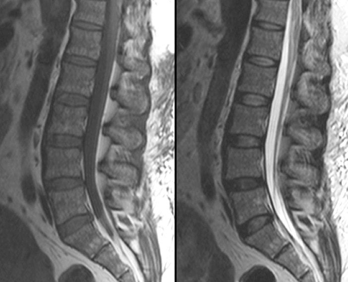
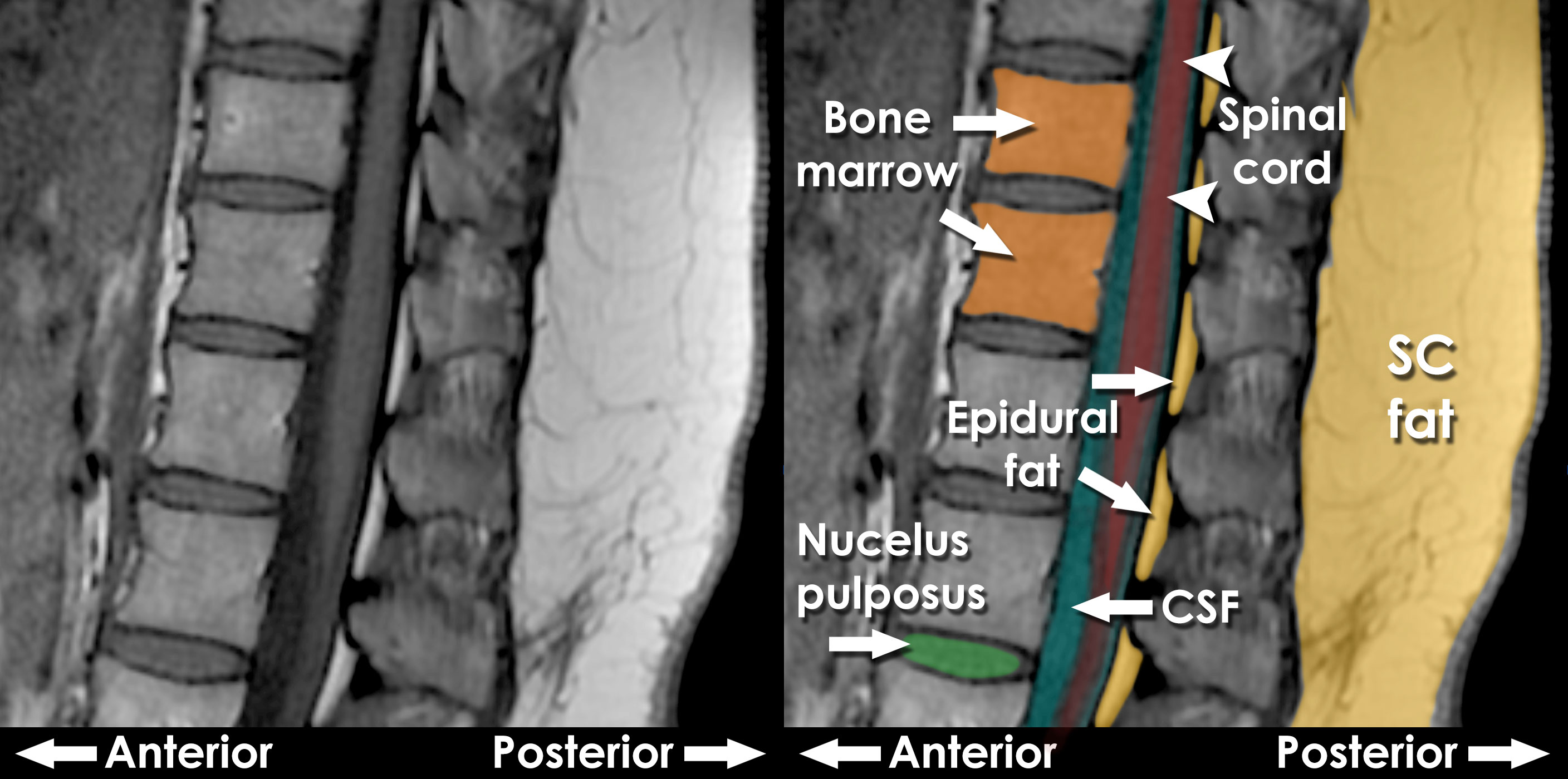
Canon Medical Systems Corporation - Check out these high-definition T1 and  T2 weighted lumbar spine images which were acquired with high  signal-to-noise ratio (#SNR) allowing #visualization of spinal cord, bone,  and disk
Canon Medical Systems Corporation - Check out these high-definition T1 and T2 weighted lumbar spine images which were acquired with high signal-to-noise ratio (#SNR) allowing #visualization of spinal cord, bone, and disk

MRI Basics
MRI Basics

xmlinkhub
xmlinkhub

Neurological Examination Spinal Cord Part 3 - Everything You Need To Know -  Dr. Nabil Ebraheim - YouTube
Neurological Examination Spinal Cord Part 3 - Everything You Need To Know - Dr. Nabil Ebraheim - YouTube
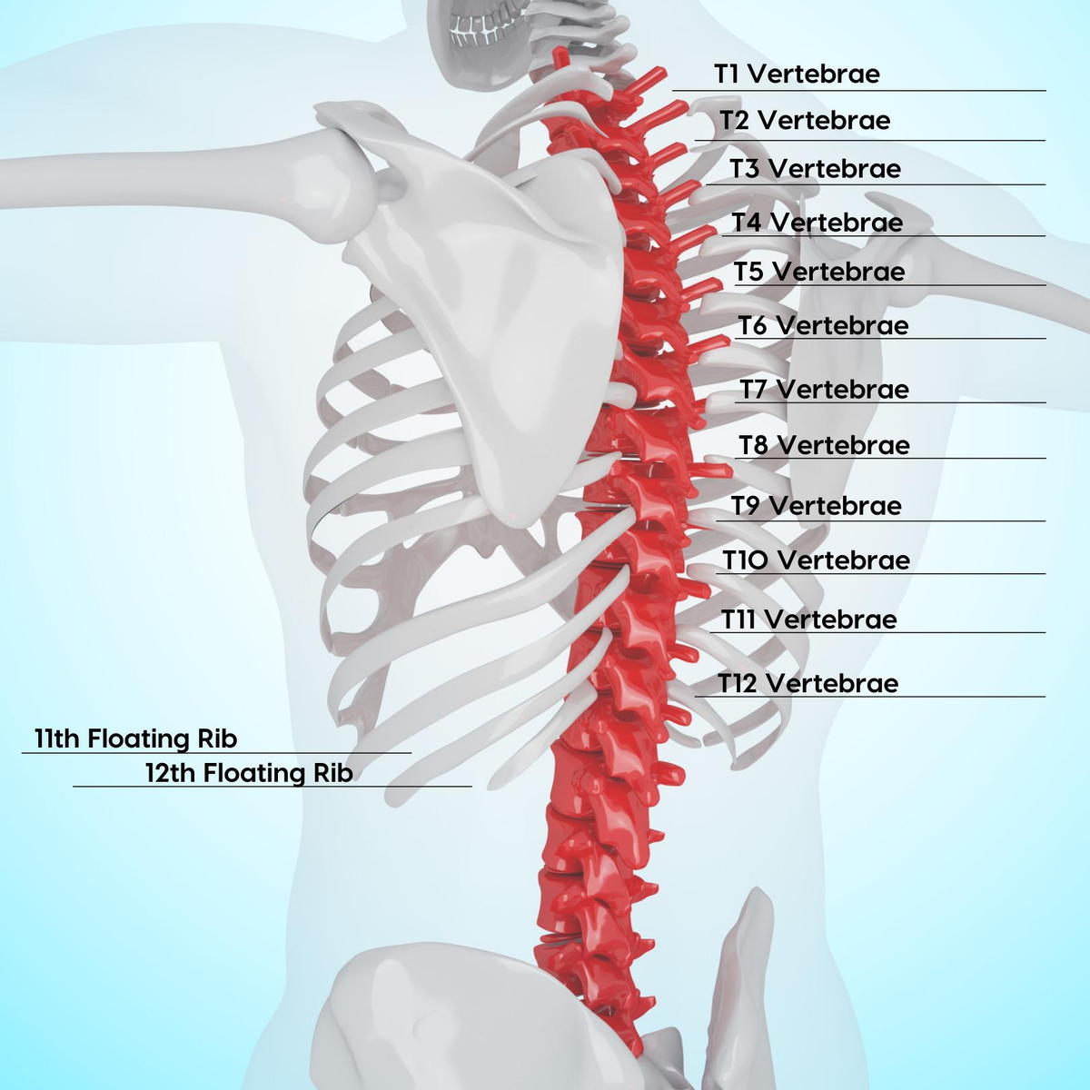
Thoracic Spine Nerves and Subluxation Gallatin Valley Chiropractic:  Bozeman, MT: Back and Neck Pain, Whiplash & More
Thoracic Spine Nerves and Subluxation Gallatin Valley Chiropractic: Bozeman, MT: Back and Neck Pain, Whiplash & More

Comparison of Sagittal FSE T2, STIR, and T1-Weighted Phase-Sensitive  Inversion Recovery in the Detection of Spinal Cord Lesions in MS at 3T |  American Journal of Neuroradiology
Comparison of Sagittal FSE T2, STIR, and T1-Weighted Phase-Sensitive Inversion Recovery in the Detection of Spinal Cord Lesions in MS at 3T | American Journal of Neuroradiology

Thoracic vertebrae: Anatomy, function and definition | Kenhub

EPOS™
EPOS™

Thoracic spondylotic myelopathy presumably caused by diffuse idiopathic  skeletal hyperostosis in a patient who underwent decompression and  percutaneous pedicle screw fixation - Shota Miyoshi, Tadao Morino, Haruhiko  Takeda, Hiroshi Nakata, Masayuki Hino,
Thoracic spondylotic myelopathy presumably caused by diffuse idiopathic skeletal hyperostosis in a patient who underwent decompression and percutaneous pedicle screw fixation - Shota Miyoshi, Tadao Morino, Haruhiko Takeda, Hiroshi Nakata, Masayuki Hino,

Normal Sagittal MRI
Normal Sagittal MRI

thoracic spine | Musculoskeletal Key

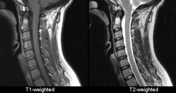
T1 vs T2 MRI | T1and T2 MRI image comparison

MRI of Spinal Bone Marrow: Part 2, T1-Weighted Imaging-Based Differential  Diagnosis | AJR
MRI of Spinal Bone Marrow: Part 2, T1-Weighted Imaging-Based Differential Diagnosis | AJR

What do T1 and T2 weighted images mean in an MRI? - Quora

Intelligent Chiropractic - T1, T2, & T3: The Top Three Thoracic Vertebrae  Tension, Tightness, & Tech Neck Posture are also 3 T's associated with  symptoms that affect your upper thoracic spine. When
Intelligent Chiropractic - T1, T2, & T3: The Top Three Thoracic Vertebrae Tension, Tightness, & Tech Neck Posture are also 3 T's associated with symptoms that affect your upper thoracic spine. When
Diagnostics | Free Full-Text | Primary Benign Neoplasms of the Spine

View Image
View Image

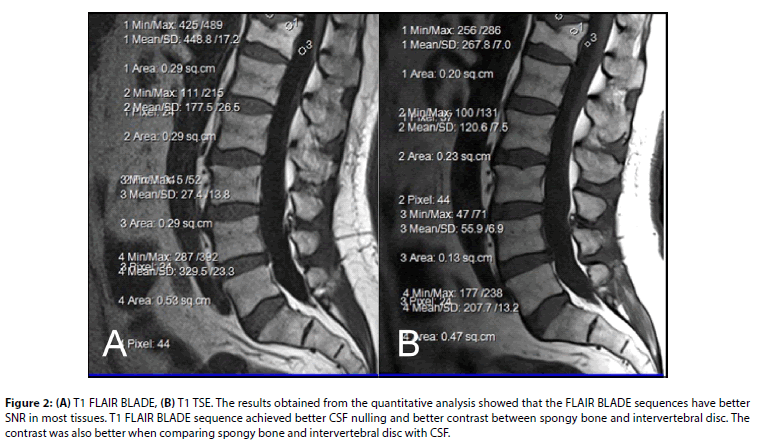
Comparison of T1 FLAIR BLADE with and without parallel imaging against T1  turbo spin echo in the MR imaging of lumbar spine in the sagittal plane
Comparison of T1 FLAIR BLADE with and without parallel imaging against T1 turbo spin echo in the MR imaging of lumbar spine in the sagittal plane

Comparing T1-weighted and T2-weighted three-point Dixon technique with  conventional T1-weighted fat-saturation and short-tau inversion recovery  (STIR) techniques for the study of the lumbar spine in a short-bore MRI  machine - ScienceDirect
Comparing T1-weighted and T2-weighted three-point Dixon technique with conventional T1-weighted fat-saturation and short-tau inversion recovery (STIR) techniques for the study of the lumbar spine in a short-bore MRI machine - ScienceDirect
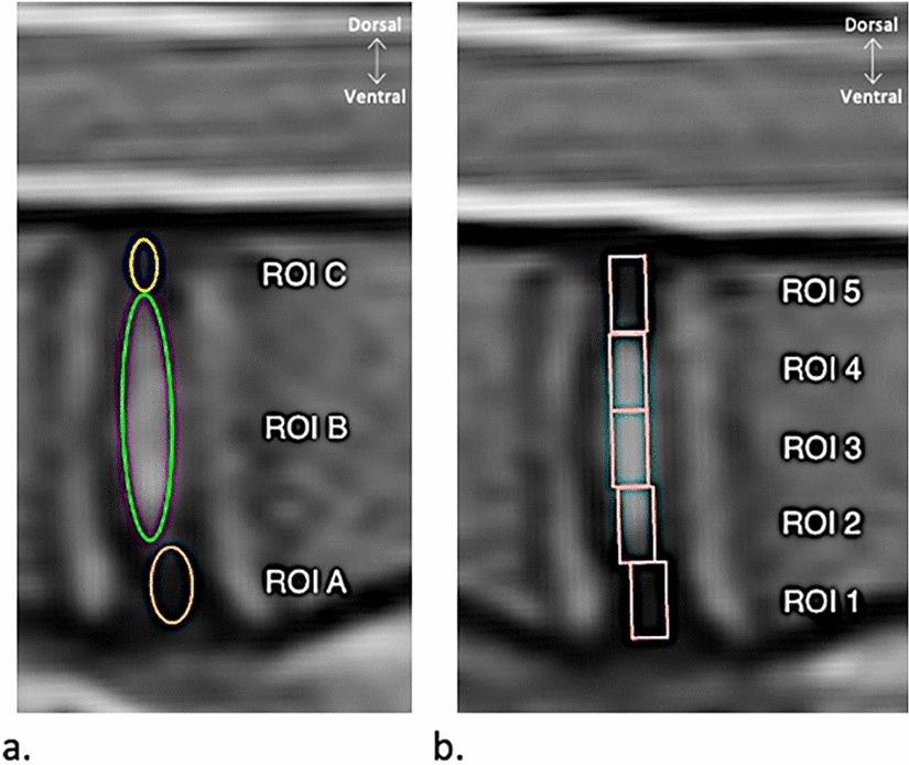
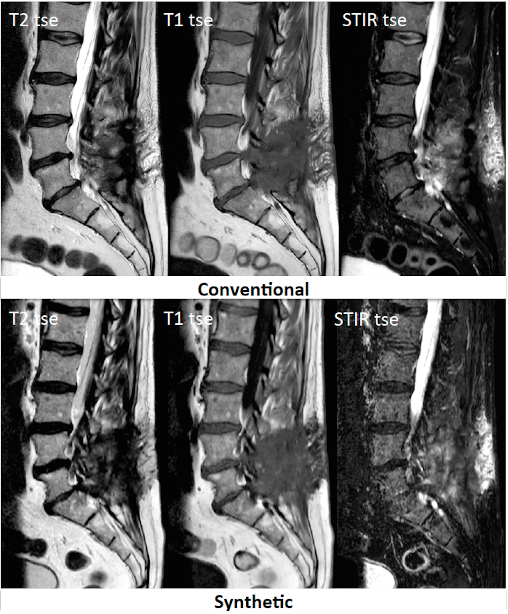
Feasibility of Synthetic MRI of the spine – fast and quantitative imaging

Comparison of MRI T1, T2, and T2* mapping with histology for assessment of  intervertebral disc degeneration in an ovine model | Scientific Reports
Comparison of MRI T1, T2, and T2* mapping with histology for assessment of intervertebral disc degeneration in an ovine model | Scientific Reports

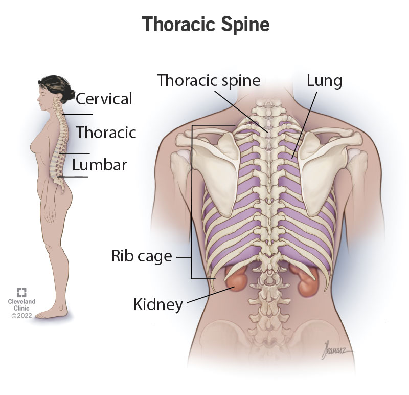
Thoracic Spine: What It Is, Function & Anatomy

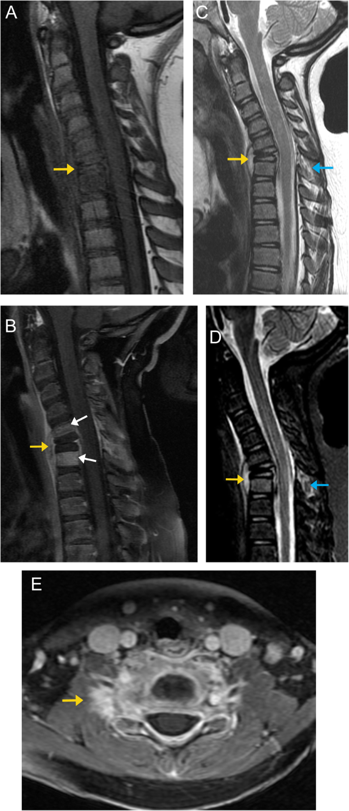
Bone up on spinal osseous lesions: a case review series | Insights into  Imaging | Full Text
Bone up on spinal osseous lesions: a case review series | Insights into Imaging | Full Text

Preoperative MRI of thoracic spine: T2 (A) and T1-weigh | Open-i

Sagittal T1-and T2-weighted image of lumbar and thoracic spine. A.... |  Download Scientific Diagram
Sagittal T1-and T2-weighted image of lumbar and thoracic spine. A.... | Download Scientific Diagram

MRI interpretation - T1 v T2 images